Morphometric Statistical Method for the Early Detection of Brain Degeneration in MR Imaging
M. Bach Cuadra, B. Mortamet, F. Budin, D. Van De Ville, R. Meuli, J.-P. Thiran
Proceedings of the CHUV Research Day (CHUV'07), Lausanne VD, Swiss Confederation, February 1, 2007, pp. 148.
Advances in neuroscience and neuroimaging have led to an increasing recognition that certain neuroanatomical structures may be affected preferentially by particular diseases. The underlying pathology is reflected through the distribution of the structural changes that may determine the clinical phenomenology. To improve diagnosis and treatment, it may be extremely useful to detect these signs as early as possible. In this domain, morphological brain studies can play a major role. Indeed, an accurate assessment of brain degeneration can rely on the high quality and resolution of modern medical imaging techniques, namely Magnetic Resonance Imaging (MRI), but requires appropriate image processing techniques to detect, locate and quantify tissue loss at an early stage. Rates of brain-tissue loss might help to distinguish between normal aging and pathological brain degeneration and lead to early diagnosis and thus treatment. Additionally, these techniques are increasingly recognized as highly necessary for the quantitative and objective assessment of drug efficacy, including the qualification of new drugs.
MRI based morphometry has experienced a remarkable gain of attention over the past few years in medical research, with one of the most prominent areas of application being the detection of statistically significant morphological differences in the brain between a population known to be affected by a certain disease and supposedly healthy controls. But one of the main limitations of this technique is the fact that, for statistical reasons, it can only compare one group against another. This is not appropriate for comparing a single patient to a control (normal) group, as we would need for early diagnosis purposes. Moreover, we have to cope with the problem of distinguishing normal aging and pathologic degeneration.
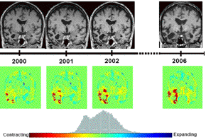
We propose a single-subject voxel-based morphometry (VBM) to guide the detection of cerebral structure abnormalities and tissue loss. While algorithms to detect group differences at one point in time (cross-sectional studies) or over time (longitudinal studies) are well known, the assessment of individual cases is more difficult but nevertheless an issue of great interest to improve clinical diagnosis. The key problem is that a number of assumptions need to hold in order for VBM to be valid and that are not completely fulfilled in the single-case vs control group. To evaluate these limitations, we propose a preliminary study where single subject brain degeneration is estimated in terms of percentiles of the distribution of normal brains. We illustrate this method on a patient diagnosed with progressive aphasia (see on Fig. 1). A group of controls is matched in age and sex with the patient (female, 60 years old). The results show significant differences in the temporal region. It is noteworthy that significant differences have been revealed already on earliest images, while they were barely visible by visual inspection.

@INPROCEEDINGS(http://bigwww.epfl.ch/publications/bachcuadra0701.html,
AUTHOR="Bach Cuadra, M. and Mortamet, B. and Budin, F. and Van De Ville,
	D. and Meuli, R. and Thiran, J.-P.",
TITLE="Morphometric Statistical Method for the Early Detection of Brain
	Degeneration in {MR} Imaging",
BOOKTITLE="CHUV Research Day ({CHUV'07})",
YEAR="2007",
editor="",
volume="",
series="",
pages="148",
address="Lausanne VD, Swiss Confederation",
month="February 1,",
organization="",
publisher="",
note="")