Biomedical Imaging Group
Logo EPFL
    • Splines Tutorials
    • Splines Art Gallery
    • Wavelets Tutorials
    • Image denoising
    • ERC project: FUN-SP
    • Sparse Processes - Book Preview
    • ERC project: GlobalBioIm
    • The colored revolution of bioimaging
    • Deconvolution
    • SMLM
    • One-World Seminars: Representer theorems
    • A Unifying Representer Theorem
Follow us on Twitter.
Join our Github.
Masquer le formulaire de recherche
Menu
BIOMEDICAL IMAGING GROUP (BIG)
Laboratoire d'imagerie biomédicale (LIB)
  1. School of Engineering STI
  2. Institute IEM
  3.  LIB
  4.  Student Projects
  • Laboratory
    • Laboratory
    • Laboratory
    • People
    • Jobs and Trainees
    • News
    • Events
    • Seminars
    • Resources (intranet)
    • Twitter
  • Research
    • Research
    • Researchs
    • Research Topics
    • Talks, Tutorials, and Reviews
  • Publications
    • Publications
    • Publications
    • Database of Publications
    • Talks, Tutorials, and Reviews
    • EPFL Infoscience
  • Code
    • Code
    • Code
    • Demos
    • Download Algorithms
    • Github
  • Teaching
    • Teaching
    • Teaching
    • Courses
    • Student projects
  • Splines
    • Teaching
    • Teaching
    • Splines Tutorials
    • Splines Art Gallery
    • Wavelets Tutorials
    • Image denoising
  • Sparsity
    • Teaching
    • Teaching
    • ERC project: FUN-SP
    • Sparse Processes - Book Preview
  • Imaging
    • Teaching
    • Teaching
    • ERC project: GlobalBioIm
    • The colored revolution of bioimaging
    • Deconvolution
    • SMLM
  • Machine Learning
    • Teaching
    • Teaching
    • One-World Seminars: Representer theorems
    • A Unifying Representer Theorem

Students Projects

Proposals  On-Going  Completed  

Estimation of the 3D structure in super-resolution fluorescence microscopy (PALM)

Spring 2011
Master Semester Project
Project: 00212

00212
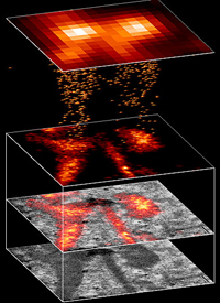
The goal of this project is to extract the 3D structure of a biological sample that has been acquired by the bi-plane PALM (Photo-Activated localization Microscopy) method. At first, a theoretical model of the PSF (Point Spread Function) will be compared with experimental data of single beads. The experimental data will then be used for calibrating the two focal planes. The third stage would consist of deriving a 3D reconstruction algorithm that will be implemented in Matlab or in Java. The algorithm will be verified by both simulated and real data.
Collaboration: Prof Suliana Manley, LEB, EPFL
  • Supervisors
  • Hagai Kirshner, hagai.kirshner@epfl.ch, 31136, BM 4.142
  • Michael Unser, michael.unser@epfl.ch, 021 693 51 75, BM 4.136
  • Daniel Sage (BM 4.135) tel 351 89
  • Laboratory
  • Research
  • Publications
  • Code
  • Teaching
    • Courses
    • Student projects
Logo EPFL, Ecole polytechnique fédérale de Lausanne
Emergencies: +41 21 693 3000 Services and resources Contact Map Webmaster email

Follow EPFL on social media

Follow us on Facebook. Follow us on Twitter. Follow us on Instagram. Follow us on Youtube. Follow us on LinkedIn.
Accessibility Disclaimer Privacy policy

© 2023 EPFL, all rights reserved