Biomedical Imaging Group
Logo EPFL
    • Splines Tutorials
    • Splines Art Gallery
    • Wavelets Tutorials
    • Image denoising
    • ERC project: FUN-SP
    • Sparse Processes - Book Preview
    • ERC project: GlobalBioIm
    • The colored revolution of bioimaging
    • Deconvolution
    • SMLM
    • One-World Seminars: Representer theorems
    • A Unifying Representer Theorem
Follow us on Twitter.
Join our Github.
Masquer le formulaire de recherche
Menu
BIOMEDICAL IMAGING GROUP (BIG)
Laboratoire d'imagerie biomédicale (LIB)
  1. School of Engineering STI
  2. Institute IEM
  3.  LIB
  4.  Student Projects
  • Laboratory
    • Laboratory
    • Laboratory
    • People
    • Jobs and Trainees
    • News
    • Events
    • Seminars
    • Resources (intranet)
    • Twitter
  • Research
    • Research
    • Researchs
    • Research Topics
    • Talks, Tutorials, and Reviews
  • Publications
    • Publications
    • Publications
    • Database of Publications
    • Talks, Tutorials, and Reviews
    • EPFL Infoscience
  • Code
    • Code
    • Code
    • Demos
    • Download Algorithms
    • Github
  • Teaching
    • Teaching
    • Teaching
    • Courses
    • Student projects
  • Splines
    • Teaching
    • Teaching
    • Splines Tutorials
    • Splines Art Gallery
    • Wavelets Tutorials
    • Image denoising
  • Sparsity
    • Teaching
    • Teaching
    • ERC project: FUN-SP
    • Sparse Processes - Book Preview
  • Imaging
    • Teaching
    • Teaching
    • ERC project: GlobalBioIm
    • The colored revolution of bioimaging
    • Deconvolution
    • SMLM
  • Machine Learning
    • Teaching
    • Teaching
    • One-World Seminars: Representer theorems
    • A Unifying Representer Theorem

Students Projects

Proposals  On-Going  Completed  

Image Analysis of the Morphology of a Spermatozoon

Autumn 2011
Master Semester Project
Project: 00219

00219
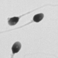
A wide corpus of microscopic images of Swiss conscripts' spermatozoa has been collected. This allows for the establishment of standards for the statistical distribution of characteristics of the spermatozoa of healthy young men. The purpose of this project is to develop software tools to which the tedious task of actually measuring the morphology of each spermatozoon can be delegated. After a spermatozoon has been automatically detected and isolated in an image, it is desired to automatically extract characteristics such as the width and length of the main axes of the head, departure of its shape from that of an ellipse, area of the head, area of a piece of the head known as the acrosome, number of vacuoles in the head, number of tails, width of the part of the tail near the head of the spermatozoon, and angle between the tail and the main axis of the head. The software tools to develop will include advanced imaging filters that take advantage of steerability to detect the structures of interest.

This programming project in Java for ImageJ is a collaboration with the Biomedical Imaging Group, the Centre hospitalier universitaire vaudois, and Biophos AG. A prerequisite is to follow (or to have followed) the course on image processing taught by M. Unser.
  • Supervisors
  • Nicolas Chenouard, nicolas.chenouard@epfl.ch, 351 43, BM 4.141
  • Michael Unser, michael.unser@epfl.ch, 021 693 51 75, BM 4.136
  • Laboratory
  • Research
  • Publications
  • Code
  • Teaching
    • Courses
    • Student projects
Logo EPFL, Ecole polytechnique fédérale de Lausanne
Emergencies: +41 21 693 3000 Services and resources Contact Map Webmaster email

Follow EPFL on social media

Follow us on Facebook. Follow us on Twitter. Follow us on Instagram. Follow us on Youtube. Follow us on LinkedIn.
Accessibility Disclaimer Privacy policy

© 2023 EPFL, all rights reserved