High-Throughput 3D Segmentation of Living Tissue
Spring 2013
Master Semester Project
Project: 00245
Thanks to substantial improvements in optics, imaging sensors, and florescence labeling methods, microscopy has matured to the point that it enables sensitive time-lapse imaging of 3D cells in vivo. One of the major challenges of current biomedical research is to characterize not just the spatial organization of these biological complex systems, but their spatio-temporal relationships as well.
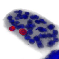
This project addresses the problem of detecting and segmenting a large number of three-dimensional biological objects and tracking them over time. This is far from trivial due to the fact that, in biology, the objects of interest are usually indistinguishable from each other and can appear tightly packed and in various configurations.
The students will first get trained on state-of-the-art 3D segmentation techniques. Then they will design and implement a software solution that they will need for analysing data acquired at the EPFL's Bio-Imaging and Optics Platform.
- Supervisors
- Ricard Delgado-Gonzalo, ricard.delgado@epfl.ch, 021 693 51 43, BM 4.141
- Michael Unser, michael.unser@epfl.ch, 021 693 51 75, BM 4.136