Biomedical Imaging Group
Logo EPFL
    • Splines Tutorials
    • Splines Art Gallery
    • Wavelets Tutorials
    • Image denoising
    • ERC project: FUN-SP
    • Sparse Processes - Book Preview
    • ERC project: GlobalBioIm
    • The colored revolution of bioimaging
    • Deconvolution
    • SMLM
    • One-World Seminars: Representer theorems
    • A Unifying Representer Theorem
Follow us on Twitter.
Join our Github.
Masquer le formulaire de recherche
Menu
BIOMEDICAL IMAGING GROUP (BIG)
Laboratoire d'imagerie biomédicale (LIB)
  1. School of Engineering STI
  2. Institute IEM
  3.  LIB
  4.  Student Projects
  • Laboratory
    • Laboratory
    • Laboratory
    • People
    • Jobs and Trainees
    • News
    • Events
    • Seminars
    • Resources (intranet)
    • Twitter
  • Research
    • Research
    • Researchs
    • Research Topics
    • Talks, Tutorials, and Reviews
  • Publications
    • Publications
    • Publications
    • Database of Publications
    • Talks, Tutorials, and Reviews
    • EPFL Infoscience
  • Code
    • Code
    • Code
    • Demos
    • Download Algorithms
    • Github
  • Teaching
    • Teaching
    • Teaching
    • Courses
    • Student projects
  • Splines
    • Teaching
    • Teaching
    • Splines Tutorials
    • Splines Art Gallery
    • Wavelets Tutorials
    • Image denoising
  • Sparsity
    • Teaching
    • Teaching
    • ERC project: FUN-SP
    • Sparse Processes - Book Preview
  • Imaging
    • Teaching
    • Teaching
    • ERC project: GlobalBioIm
    • The colored revolution of bioimaging
    • Deconvolution
    • SMLM
  • Machine Learning
    • Teaching
    • Teaching
    • One-World Seminars: Representer theorems
    • A Unifying Representer Theorem

Students Projects

Proposals  On-Going  Completed  

The double helix PSF: implementation and localization applications

Spring 2013
Master Semester Project
Project: 00253

00253
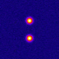
Photo-activated Localization Microscopy (PALM) provides an attractive framework for studying the living cell at a nanometer scale. It is based on exciting a sparse set of fluorophores at each time frame, and on fitting the image of each fluorophore with a point-spread function (PSF). In order to improve the axial localization accuracy of this method, one can design a PSF that exhibits larger sensitivity to the depth value. One such design is the double helix PSF, which consists of two lobes that rotate as a function of the point-source depth value. The purpose of this project is to investigate the properties of the double-helix PSF within the context of localization microscopy. The main stages of the project are: numerical optimization for the phase function that leads to the double helix pattern, implementation of the PSF model in Java, and 3-D localization analysis for different acquisition conditions: sub-pixel positions, depth values, refractive index mismatch, and noise. Background in optics and programming is recommended.
  • Supervisors
  • Hagai Kirshner, hagai.kirshner@epfl.ch, 31136, BM 4.142
  • Michael Unser, michael.unser@epfl.ch, 021 693 51 75, BM 4.136
  • Laboratory
  • Research
  • Publications
  • Code
  • Teaching
    • Courses
    • Student projects
Logo EPFL, Ecole polytechnique fédérale de Lausanne
Emergencies: +41 21 693 3000 Services and resources Contact Map Webmaster email

Follow EPFL on social media

Follow us on Facebook. Follow us on Twitter. Follow us on Instagram. Follow us on Youtube. Follow us on LinkedIn.
Accessibility Disclaimer Privacy policy

© 2023 EPFL, all rights reserved