Assessment of imaging performance in confocal fluorescence microscopy
Spring 2013
Project: 00265
The goal of this project is to conduct an experimental and theoretical study of image resolution and noise in confocal fluorescence microscopy.
The students will first get trained on modern confocal microscopes equipped with state-of-the-art detectors at EPFL's Bio-Imaging and Optics Platform. They will then acquire images of samples with nanometer-sized features---such as fluorescent beads and biological structures---while varying system parameters such as the pinhole size or the exposure time. They will have to design and apply quantitative criteria that demonstrate the compromise between image resolution and noise.
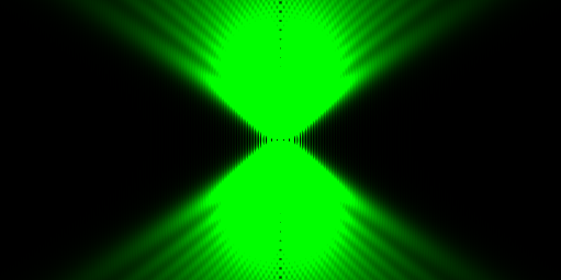
In parallel, the students will have to familiarize themselves with the physical principles of confocal microscopy. In particular, they should understand and implement (as an ImageJ plugin) a model for confocal Point-Spread Functions based on Fourier optics. As a final step, they should compare their numerical prediction with the real PSFs obtained from fluorescent beads.
The students will first get trained on modern confocal microscopes equipped with state-of-the-art detectors at EPFL's Bio-Imaging and Optics Platform. They will then acquire images of samples with nanometer-sized features---such as fluorescent beads and biological structures---while varying system parameters such as the pinhole size or the exposure time. They will have to design and apply quantitative criteria that demonstrate the compromise between image resolution and noise.
In parallel, the students will have to familiarize themselves with the physical principles of confocal microscopy. In particular, they should understand and implement (as an ImageJ plugin) a model for confocal Point-Spread Functions based on Fourier optics. As a final step, they should compare their numerical prediction with the real PSFs obtained from fluorescent beads.
- Supervisors
- Cédric Vonesch, cedric.vonesch@epfl.ch, 021 693 51 43, BM 4.141
- Michael Unser, michael.unser@epfl.ch, 021 693 51 75, BM 4.136