Biomedical Imaging Group
Logo EPFL
    • Splines Tutorials
    • Splines Art Gallery
    • Wavelets Tutorials
    • Image denoising
    • ERC project: FUN-SP
    • Sparse Processes - Book Preview
    • ERC project: GlobalBioIm
    • The colored revolution of bioimaging
    • Deconvolution
    • SMLM
    • One-World Seminars: Representer theorems
    • A Unifying Representer Theorem
Follow us on Twitter.
Join our Github.
Masquer le formulaire de recherche
Menu
BIOMEDICAL IMAGING GROUP (BIG)
Laboratoire d'imagerie biomédicale (LIB)
  1. School of Engineering STI
  2. Institute IEM
  3.  LIB
  4.  Student Projects
  • Laboratory
    • Laboratory
    • Laboratory
    • People
    • Jobs and Trainees
    • News
    • Events
    • Seminars
    • Resources (intranet)
    • Twitter
  • Research
    • Research
    • Researchs
    • Research Topics
    • Talks, Tutorials, and Reviews
  • Publications
    • Publications
    • Publications
    • Database of Publications
    • Talks, Tutorials, and Reviews
    • EPFL Infoscience
  • Code
    • Code
    • Code
    • Demos
    • Download Algorithms
    • Github
  • Teaching
    • Teaching
    • Teaching
    • Courses
    • Student projects
  • Splines
    • Teaching
    • Teaching
    • Splines Tutorials
    • Splines Art Gallery
    • Wavelets Tutorials
    • Image denoising
  • Sparsity
    • Teaching
    • Teaching
    • ERC project: FUN-SP
    • Sparse Processes - Book Preview
  • Imaging
    • Teaching
    • Teaching
    • ERC project: GlobalBioIm
    • The colored revolution of bioimaging
    • Deconvolution
    • SMLM
  • Machine Learning
    • Teaching
    • Teaching
    • One-World Seminars: Representer theorems
    • A Unifying Representer Theorem

Students Projects

Proposals  On-Going  Completed  

Region of Interest Computed Tomography

Spring 2017
Master Semester Project
Project: 00338

00338
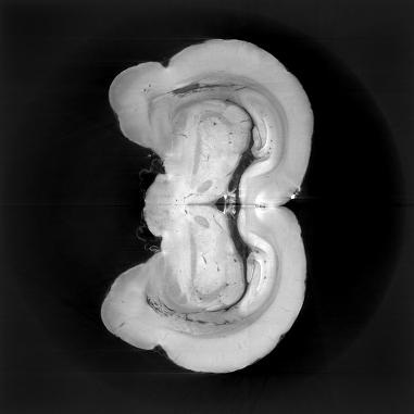
In this project, we aim to develop a method for region of interest (ROI) computed tomography (CT) reconstruction. In X-ray CT, the images of slices of an object are reconstructed from a set of X-ray images of the object taken from different angles. This technique has found applications in a broad range of areas including materials science engineering, archaeology, biology, and medicine. Especially in biomedical applications, the user is often interested only in a small ROI inside a larger volume, however, it is non-trivial in CT to reconstruct anything but the entire field of view because objects outside the ROI cause artifacts in the ROI reconstruction. We want to explore ways of creating a high-resolution reconstruction of an ROI by using a low-resolution full field of view reconstruction to correct for these artifacts.
  • Supervisors
  • Mike McCann, michael.mccann@epfl.ch, BM 4141
  • Michael Unser, michael.unser@epfl.ch, 021 693 51 75, BM 4.136
  • Laboratory
  • Research
  • Publications
  • Code
  • Teaching
    • Courses
    • Student projects
Logo EPFL, Ecole polytechnique fédérale de Lausanne
Emergencies: +41 21 693 3000 Services and resources Contact Map Webmaster email

Follow EPFL on social media

Follow us on Facebook. Follow us on Twitter. Follow us on Instagram. Follow us on Youtube. Follow us on LinkedIn.
Accessibility Disclaimer Privacy policy

© 2023 EPFL, all rights reserved