| Mourad Kharouf | Semester project |
| Section Microtechnique, EPFL | February 1999 |
In collaboration with: Hôpital Ophtalmologique Jules Gonin de Lausanne
The retina is the noble part of the human eye. The macula is situated in the centre of the retina. This region is the most important since it controls the vision. For elder people, we can observe sometimes leaking around the macula, this can cause the legal blindness after two years for a non treated patient. The macula degeneration affects more than 500'000 people in the industrial countries each year. For the past five years, many studies have been undertaken to cure this disease.
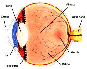
Fig. 1 Position of the macula
The doctor injects a fluorescent drug which reacts, when excited by a laser beam, to kill the neighbour cells. However, he has to be sure that it does not harm the photoreceptors. The diffusion of the drug can be seen by a camera and it will be concentrated in the leaking regions. A stack of pictures of the retina is taken during this operation. However, since the retina is always moving, it is difficult for the doctor to estimate the diffusion speed of the drug in the leaking area and to control other parameters very important for the treatment.
For the time being, the alignment of those pictures is done manually by choosing a reference point in each image and then reorganising the stack by translating them. However, this method is time consuming and not very accurate since it does not handle a general transformation between two consecutive pictures.
The aim of the project is to align the retina pictures automatically. In the first part, we will verify and improve the manual compensation of the retina motion by choosing three reference points instead of one. Then we will do some experiments and discuss the results. In the second part, we will implement a program that does the automatic compensation of the retina motion using the LsAlign program based on a Least Square criterion. The LsAlign program aligns a test image with a reference image by computing the difference between the reference image and the output of the test image by an affine transformation. The transformation that gives the minimum error will be chosen.
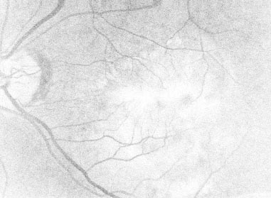
Fig. 2 Image of the retina and diffusion of the drug in the vessels
We will test this program on the original stack using all the pictures and compare the results with those of the first part.
We will propose as well different features in order to improve the performances of the automatic processing. We will implement a similar program with an automatic elimination of the bad pictures. We will do other tests using a pre-processed images in order to improve the automatic alignment between two images.