|
When looking at a three-dimensional (3D) specimen through a transmitted brightfield optical microscope, only the part of the specimen contained in the focal plane
appears sharp while the remainder of the specimen looks smooth. The deconvolution task consists in deblurring the observed image in order
to recover the original shapes of the object.
The goal of this project is the implementation of a java plug-in for the software ImageJ.
The first step is the caracterization of the point spred function (PSF) of the microscope. Since this function is the mathematical link between the
observation and the actual object, a good knowledge of the PSF is important for optimum results. A simplified model (Agard 1984) and a more accurate
one (Gibson 1991) have been developped.


(Left to right) Agard's model, measurement, Gibson's model
The figure above shows the comparison between the models and measurements. While Agard's model is symetric, Gibson's model
can generate asymetric PSFs closer to real observations.
Once the microscope has been caracterized, deconvolution algorithms can be implemented. Although 2D algorithms have been widely used,
their adaptation in 3D gives rise to many problems leading to the impossibility to recover the original image without any assumption. Moreover
some noise is added to the signal ; it has been considered as a white Gaussian noise.
The algorithms that have been implemented are : inverse filtering, regularized inverse, iterative Van Cittert's algorithm and ForWaRD
(Neelamani R. , Choi H. , Baraniuk R. 2003).
|
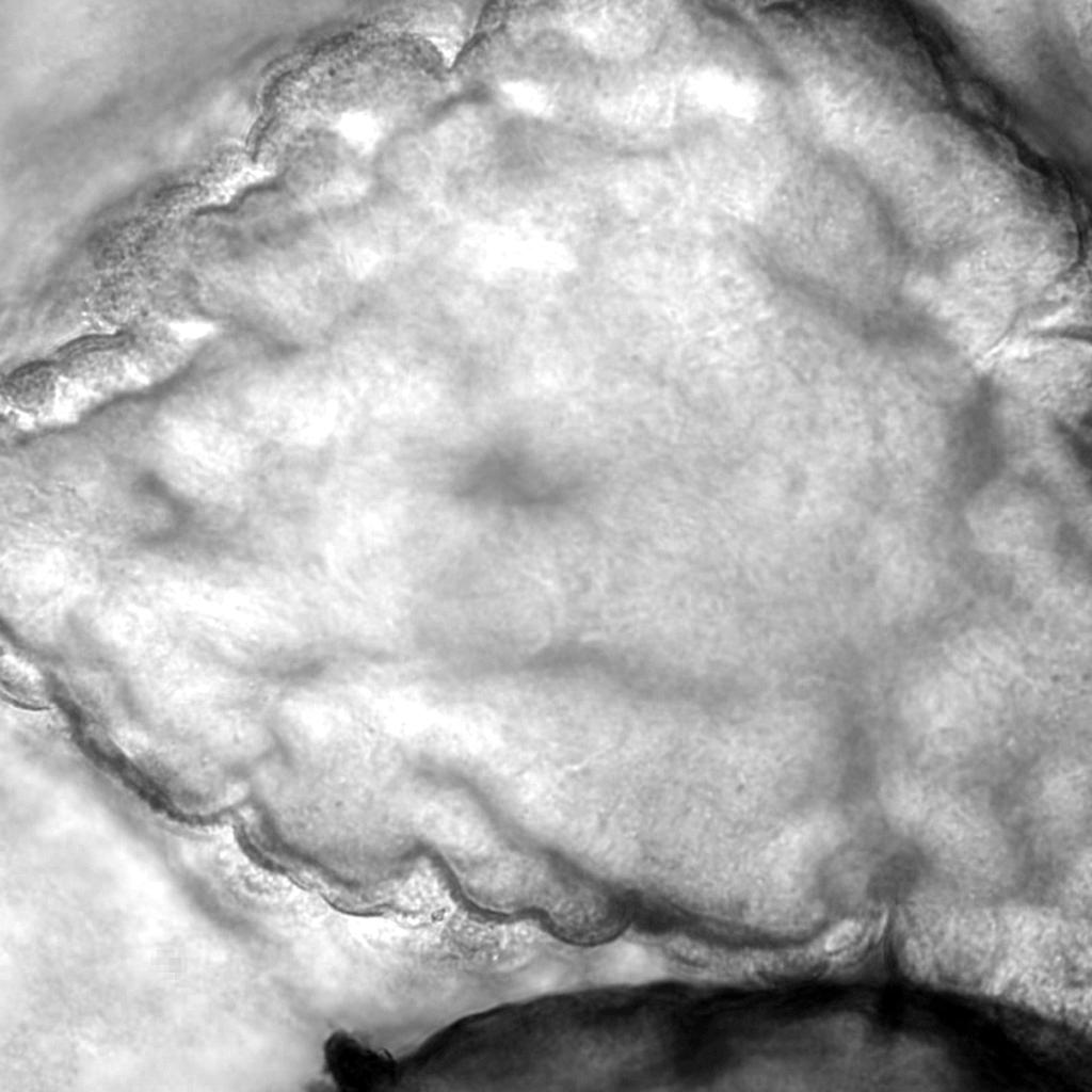
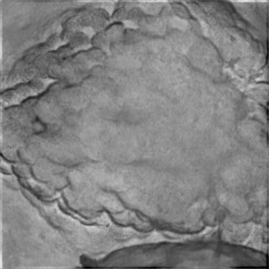
Obervation -- Regularized deconvolution
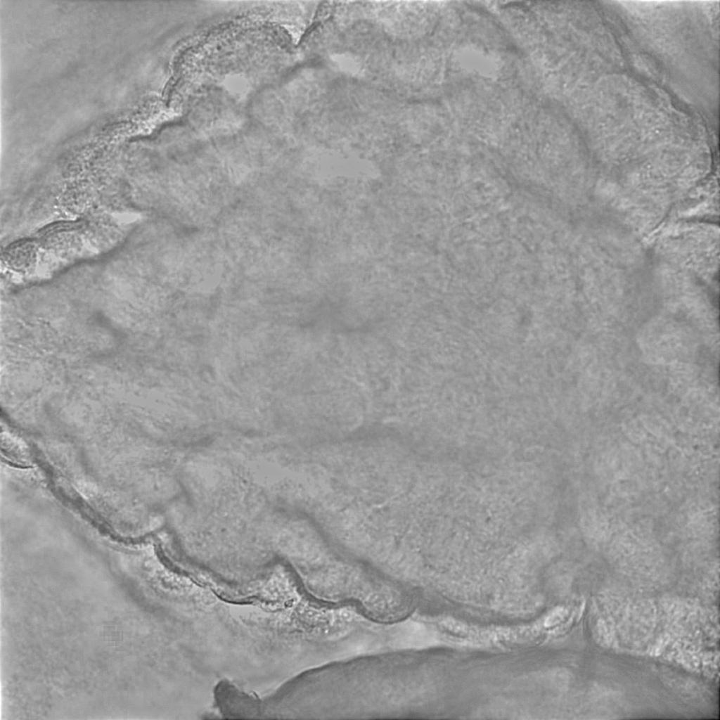
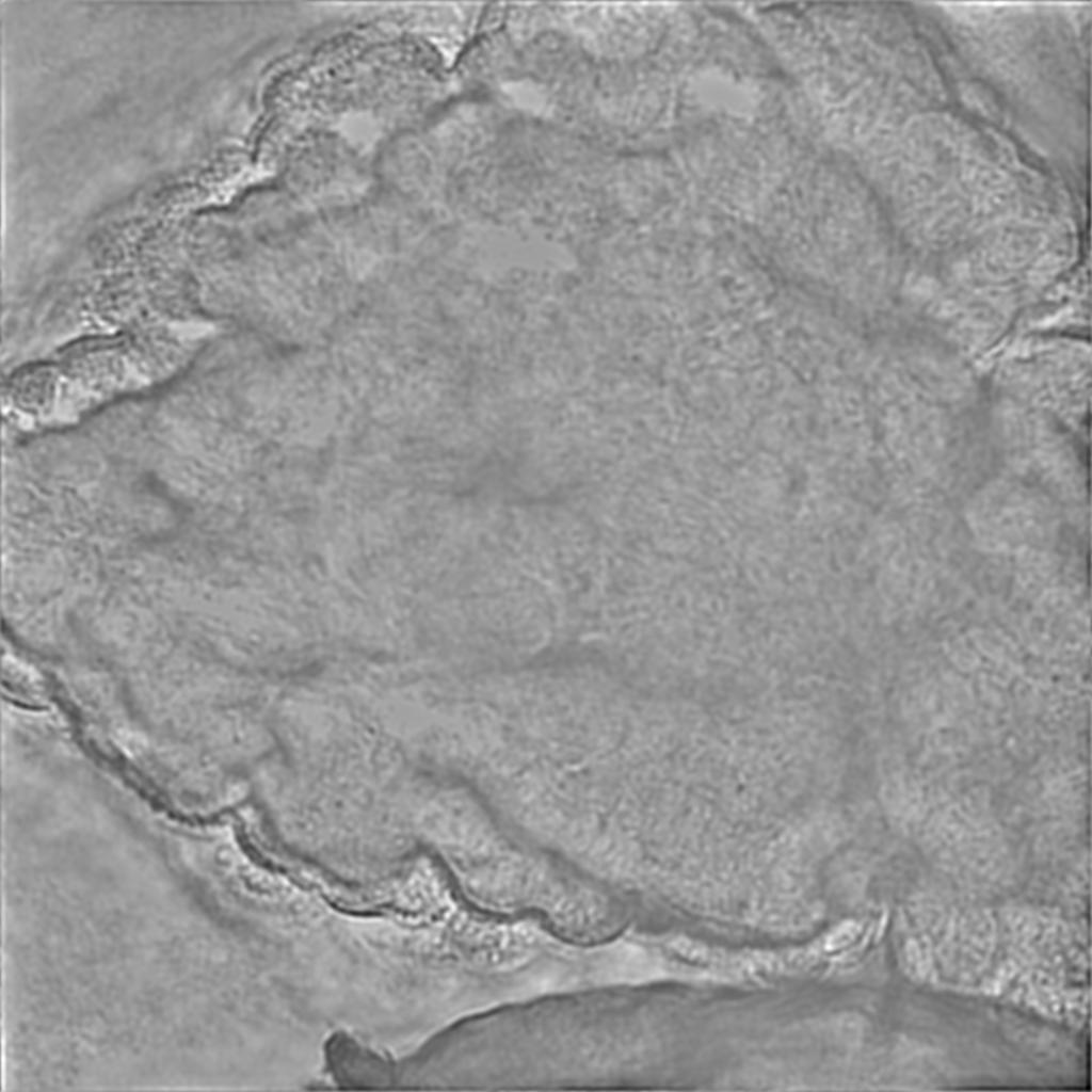
Van Cittert deconvolution (40 iterations) -- ForWaRD
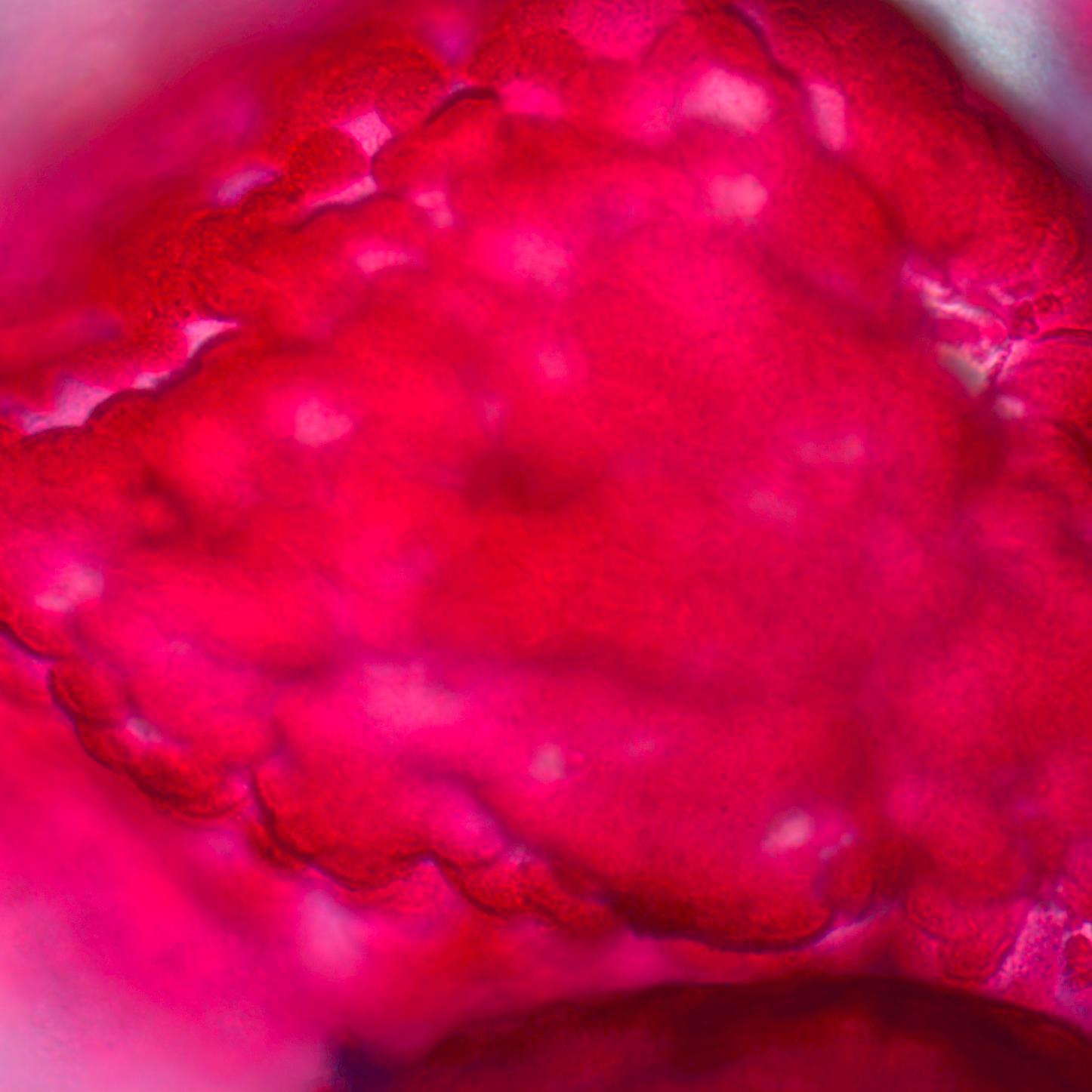
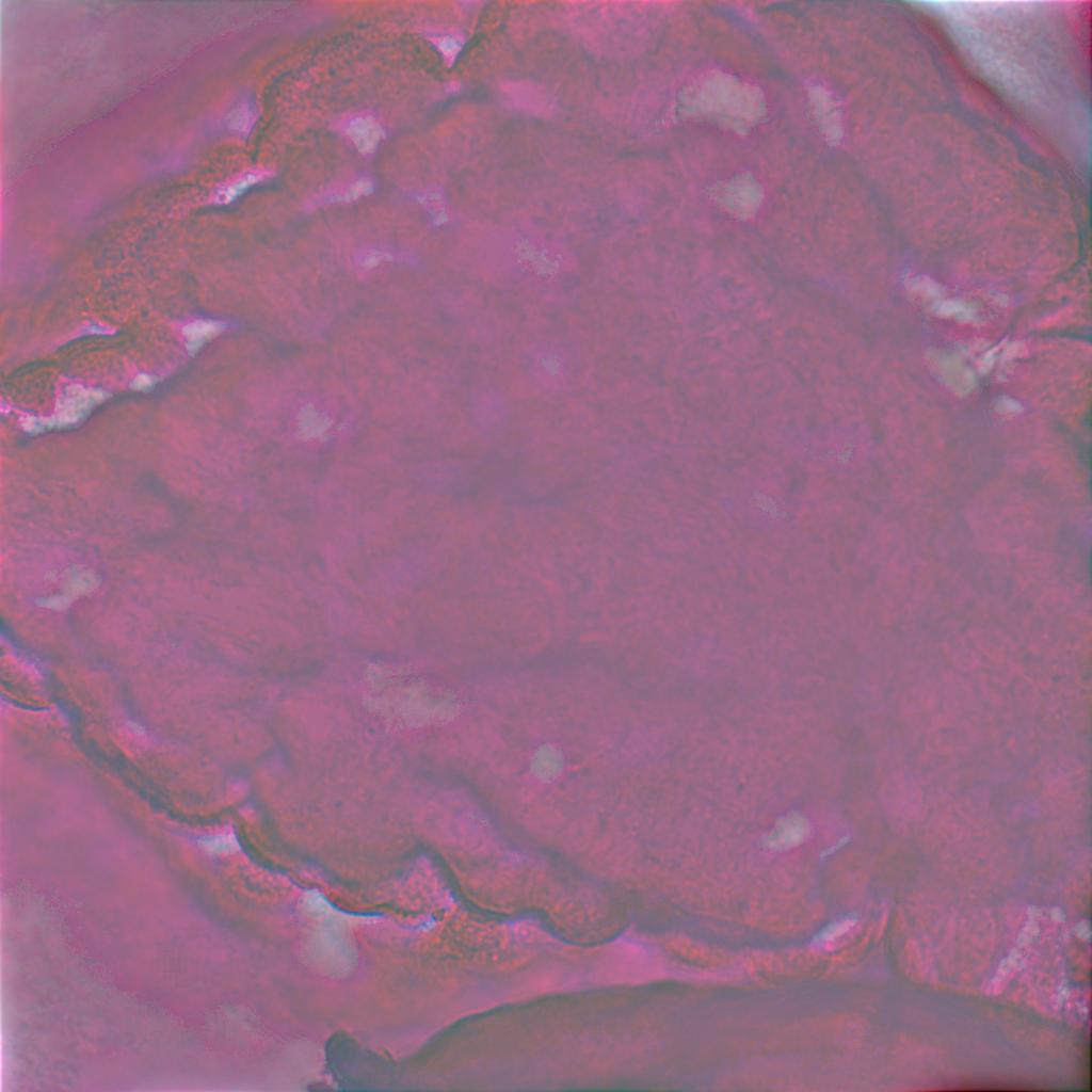
Color observation -- Color Van Cittert deconvolution
Visualization of the results
While the level of details is clearly improved, informations as the color may be lost or not recovered well. However the level of
details remains much higher.
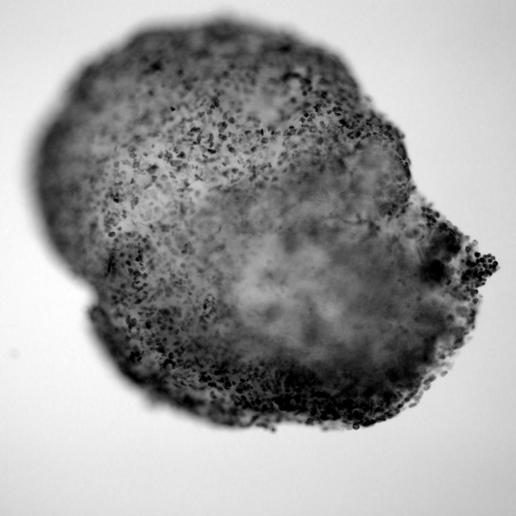
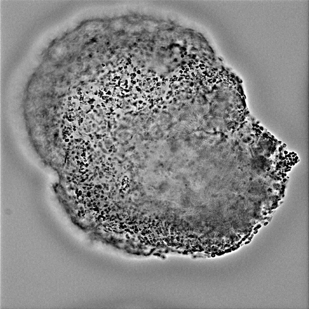
Other example : image of a pancreas and deconvolution (Van Cittert, 40 iterations)
|
