Motivation
The recent developments in biotechnology has made it necessary for the biologists to be able
to observe small biological structures such as DNA, viruses, bacteria or protein
molecules in detail. Derived from electon microscopy, cryo-electron microscopy is a recently developed
method that offers many advantages for observing such living structures. However, the very poor quality
of the images that can be obtained is a major downside of this method. In this context, image processing
can be a good alternative to enhance cryo-electron microscopy images.
Work
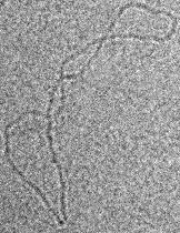
In this project we aim at reconstructing
the boundaries of DNA-molecules from such an image (Fig. 1) automatically. For this challenging task we first
use a feature detection method based on the multiscale computation of local moments. We then experiment two
contour following methods for reconstructing the DNA strand. The first one consists in selecting the most relevant structures
in a post-processing step (that involves a combination of non-maximum suppression and
hystheresis threshold algorithms), and afterwards a basic contour follwing method is applied. In the second one, a more complex algorithm
based on dynamic programming performs the contour tracking operation on a non-post-processed image.
Results
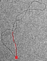
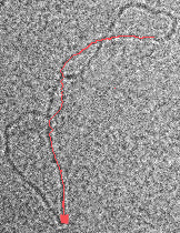
Both methods were implemented and their efficiency confirmed on synthetic images. However, It was not possible to achieve
a complete and automatic reconstruction of the DNA strand on a real image (Fig.2-3).
Some human interaction may be needed to obtain a better performance.
|