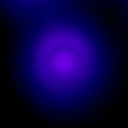
This test volume contains a fluorescent bead of known dimension—its diameter is precisely 2.5 μm. These data have the advantage of offering a simple object on which it is easy to perform a quantitative validation of the recovering of shape and dimension, before and after deconvolution.
Data
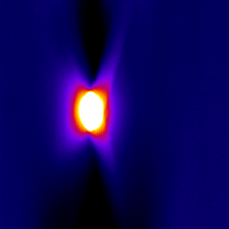
PSF
|
Image size: 256x256 pixels Number of z slices: 256 slices Number of channels: 1 Dynamic: 16 bits Image format: TIFF | |||||||||||||||||||||||||||
 XY section |
 ZY section |
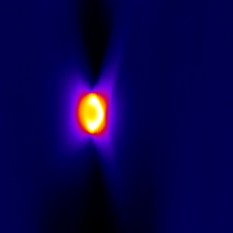 XZ section |