Geometric Alignment and Chromatic Calibration of Serial Radiographic Images
C. Dornier, L. Dorsaz-Brossa, P. Thévenaz, F. Casagni, P. Brochut, A. Mombelli, J.-P. Vallée
Dentomaxillofacial Radiology, vol. 33, no. 4, pp. 220–225, July 2004.
Objective—To develop software for automated registration and intensity calibration of serial dental radiographs for the analysis of longitudinal changes in bone density.
Methods—Serial dental radiographs were acquired using a positioning device designed to minimize projection divergence. Each radiograph included an image of a standardized aluminium wedge. The radiographs were scanned on a flatbed scanner (AGFA Duo Scan) with a spatial resolution of 300 dpi, and pixel intensity coded in 16-bit grey scale. The intensity was calibrated using serial images of selected areas with defined thickness of the aluminium wedge. A robust B-splines multiresolution registration algorithm was implemented to overcome the acquisition misalignment. Radiographs, taken before and after periodontal therapy, were subtracted to assess bone density evolution.
Results—The intensity calibration decreased the maximum intensity variations between serial radiographs from 30±17% to 1±1% (mean±standard deviation), and improved the visual comparison between the radiographs. The registration stage allowed correcting the misalignment of the radiographs on the scanner screen and superimposing the radiography contents. The observed residual motion was about 0.02±0.01 mm.
Conclusion—Very user-friendly software was developed. The manipulator needs to scan the radiographs only one time. The software performs all subsequent processing steps.
This paper is available for purchase from here.
Data Supplement—The figures are labeled according to their enumeration in the article.
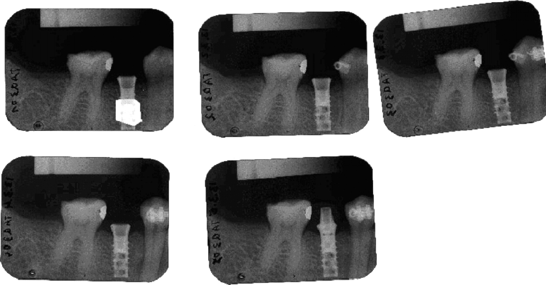
- Figure 1 (JPEG Picture) (228 kb): Example of scanned serial dental radiographs. The region of interest (ROI) (in video inverse) on the first frame represents the area used for performing the geometric registration. The ROI is always placed over the implant location.
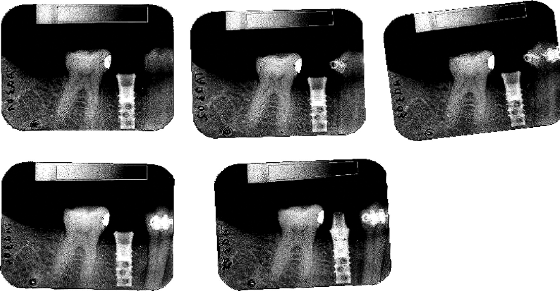
- Figure 2 (JPEG Picture) (332 kb): Definition of the region of interest (ROI) for performing the intensity calibration. A polygonal ROI was drawn over the aluminium wedge on each digital radiograph. Calibration curves are obtained by fitting the intensity profile along the rectangle length by a third order polynomial.
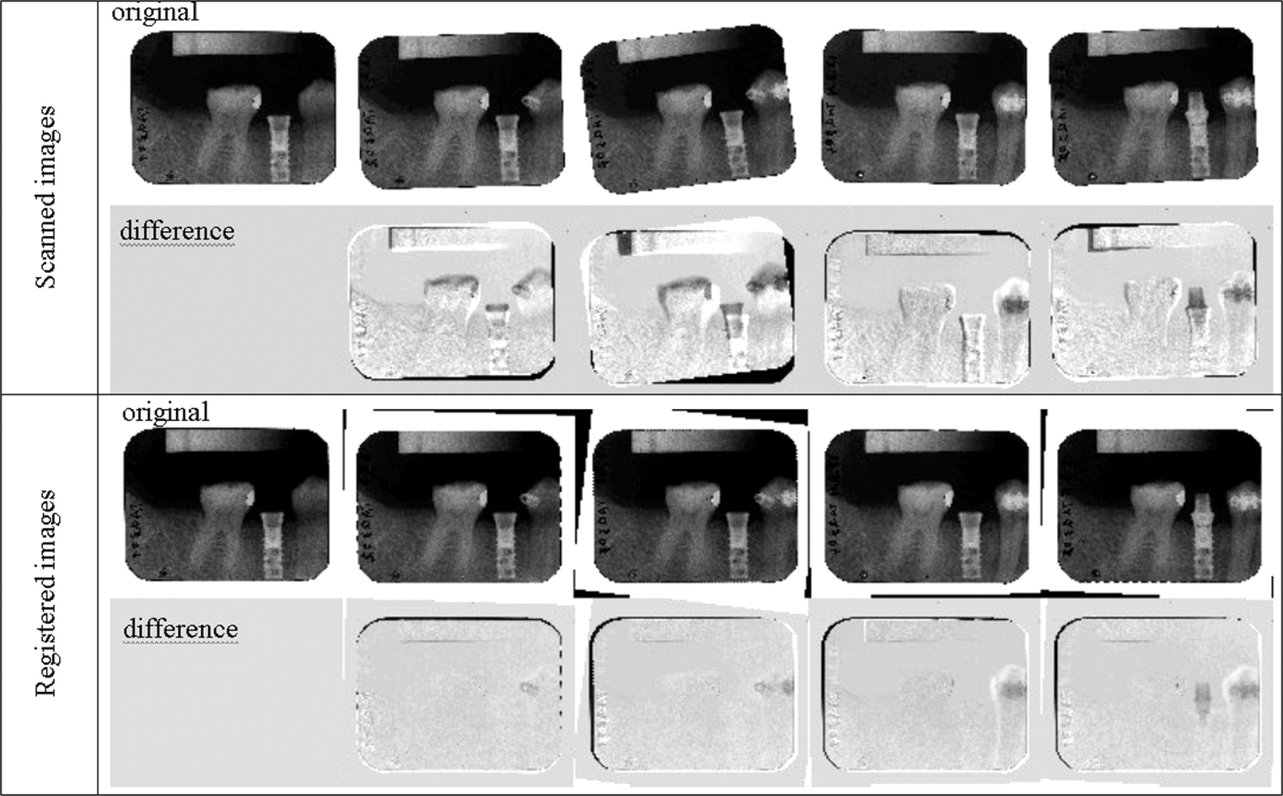
- Figure 3 (JPEG Picture) (296 kb): Example of serial dental radiographs before and after the registration process performed on the implant area. The first two lines present the scanned radiographs and the difference between each radiograph to the first radiograph. The last two lines present the same images after performing the registration stage. On difference images, the implant is clearly visible on scanned images owing to misalignment and it becomes invisible after the registration process.
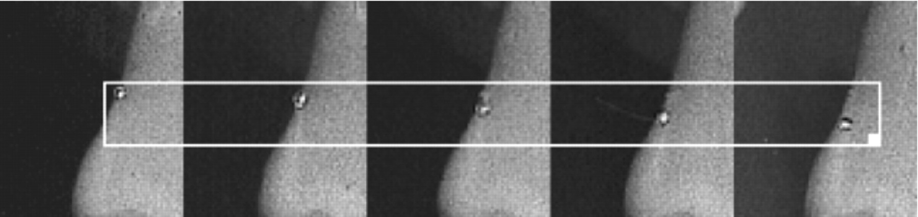
- Figure 4 (JPEG Picture) (92 kb): Serial images of the same mark performed under an optical microscope on a molar tooth. The five images were registered with the automatic algorithm. The misalignment of the mark between the different radiographs was easily observed. This example corresponded to the worst case included in this study.
@ARTICLE(http://bigwww.epfl.ch/publications/dornier0401.html,
AUTHOR="Dornier, C. and Dorsaz-Brossa, L. and Th{\'{e}}venaz, P. and
	Casagni, F. and Brochut, P. and Mombelli, A. and Vall{\'{e}}e,
	J.-P.",
TITLE="Geometric Alignment and Chromatic Calibration of Serial
	Radiographic Images",
JOURNAL="Dentomaxillofacial Radiology",
YEAR="2004",
volume="33",
number="4",
pages="220--225",
month="July",
note="")