Collagen filaments detection
Spring 2010
Master Semester Project
Project: 00193
Collagen is a protein very abundant in human body. In arterial wall, collagen serves as the main load-bearing component of the tissue, and together with elastine and smooth muscle cells, it determines its mechanical behaviour. Particularly, the spatial organization and the orientation of the filaments significantly determine tissue mechanical properties.
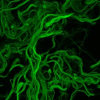
3D Images of arterial wall sections have been taken with a confocal microscope. Collagen was marked with fluorescent green staining.
The aim of the project is to segment collagen bunches of fibres so to collect information about filaments dimension and orientation for a complete understanding of collagen architecture in arterial wall. The task is to processing the images to detect filaments of different scale and different orientations using steerable filters, then a track of filament will be implemented.
The implementation of the algorithm will be done in Java as an ImageJ plugin. The project is in collaboration with the BioImaging and Optics platform of the Life Science at the EPFL.
- Supervisors
- Daniel Sage, daniel.sage@epfl.ch, 021 693 51 89, BM 4.135
- Michael Unser, michael.unser@epfl.ch, 021 693 51 75, BM 4.136
- Alessandra Griffa, BIOP, EPFL