Identification of Physical Properties Obtained at Nanometer Scale by Combining Infrared Spectroscopy with Atomic-Force Microscopy
Autumn 2014
Master Semester Project
Project: 00278
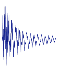
The recent technique named nanoscale infrared spectroscopy (nanoIR) uses short pulses of a tunable laser to irradiate at selected wavelengths a specimen placed under the tip of an atomic-force microscope (AFM). In the regions where light is absorbed, the abrupt raise in temperature is accompanied by an abrupt thermal expansion of the specimen. This expansion plucks the cantilever supporting the tip of the AFM. The ringing response of the cantilever contains several vibrational modes; the amplitude of each mode and its decay through time contains rich information about several properties of the specimen, which stays in contact with the tip of the AFM. This information can be recovered with unprecedented spatial resolution.
The goal of this project is to develop a signal-processing approach to extract, from the ringing response, properties such as the local coefficient of absorption of the light at each wavelength (infrared, mainly) and the local stiffness of the specimen. The measurements will need to be made robust to noise and impervious to statistical bias. The vibrational signal will be decomposed as the superposition of several vibrational modes, each with independent amplitude, phase, frequency, and decay.
Type of work: algorithmic development and programming in Java
Prerequisites: to follow or to have followed the course on signal processing taught by M. Unser
This work is in collaboration with Andrzej Kulik, Laboratoire de physique de la matière vivante, EPFL.
- Supervisors
- Philippe Thévenaz, philippe.thevenaz@epfl.ch, 021 693 51 61, BM 4.137
- Michael Unser, michael.unser@epfl.ch, 021 693 51 75, BM 4.136