Biomedical Imaging Group
Logo EPFL
    • Splines Tutorials
    • Splines Art Gallery
    • Wavelets Tutorials
    • Image denoising
    • ERC project: FUN-SP
    • Sparse Processes - Book Preview
    • ERC project: GlobalBioIm
    • The colored revolution of bioimaging
    • Deconvolution
    • SMLM
    • One-World Seminars: Representer theorems
    • A Unifying Representer Theorem
Follow us on Twitter.
Join our Github.
Masquer le formulaire de recherche
Menu
BIOMEDICAL IMAGING GROUP (BIG)
Laboratoire d'imagerie biomédicale (LIB)
  1. School of Engineering STI
  2. Institute IEM
  3.  LIB
  4.  Student Projects
  • Laboratory
    • Laboratory
    • Laboratory
    • People
    • Jobs and Trainees
    • News
    • Events
    • Seminars
    • Resources (intranet)
    • Twitter
  • Research
    • Research
    • Researchs
    • Research Topics
    • Talks, Tutorials, and Reviews
  • Publications
    • Publications
    • Publications
    • Database of Publications
    • Talks, Tutorials, and Reviews
    • EPFL Infoscience
  • Code
    • Code
    • Code
    • Demos
    • Download Algorithms
    • Github
  • Teaching
    • Teaching
    • Teaching
    • Courses
    • Student projects
  • Splines
    • Teaching
    • Teaching
    • Splines Tutorials
    • Splines Art Gallery
    • Wavelets Tutorials
    • Image denoising
  • Sparsity
    • Teaching
    • Teaching
    • ERC project: FUN-SP
    • Sparse Processes - Book Preview
  • Imaging
    • Teaching
    • Teaching
    • ERC project: GlobalBioIm
    • The colored revolution of bioimaging
    • Deconvolution
    • SMLM
  • Machine Learning
    • Teaching
    • Teaching
    • One-World Seminars: Representer theorems
    • A Unifying Representer Theorem

Students Projects

Proposals  On-Going  Completed  

Quantification of fiber-like structures in time-lapse fluorescence microscopy images

Autumn 2016
Bachelor Project
Project: 00324

00324
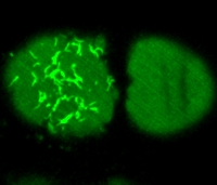
Asymmetric division is crucial for the development of multicellular organisms. The embryo of the nematode C. elegans is a well-suited model system to analyze the mechanisms of asymmetric cell division. Using spinning disc confocal microscopy, we performed live imaging of green fluorescent-tagged force generation proteins of C. elegans zygotes. This revealed that such proteins form dynamic fiber-like structures as well as foci. This student project entails designing image-analysis algorithms and applying these methods to monitor the behavior of these structures in a quantitative manner over the sequence of images. The project will be implemented in Java as a ImageJ or Icy plugin.

Collaboration with Melina Scholze, Gonczy Lab (http://gonczy-lab.epfl.ch)

  • Supervisors
  • Daniel Sage, daniel.sage@epfl.ch, 021 693 51 89, BM 4.135
  • Michael Unser, michael.unser@epfl.ch, 021 693 51 75, BM 4.136
  • Laboratory
  • Research
  • Publications
  • Code
  • Teaching
    • Courses
    • Student projects
Logo EPFL, Ecole polytechnique fédérale de Lausanne
Emergencies: +41 21 693 3000 Services and resources Contact Map Webmaster email

Follow EPFL on social media

Follow us on Facebook. Follow us on Twitter. Follow us on Instagram. Follow us on Youtube. Follow us on LinkedIn.
Accessibility Disclaimer Privacy policy

© 2023 EPFL, all rights reserved