Tracking bacterial DNA replication by quantitative analysis of the replisome foci
2022
Master Semester Project
Project: 00423
Motivation: DNA replication is one of the essential events for all kinds of life.
This project aims to utilize image processing and analysis tools to quantitatively interpret the microscopic imaging data and improve our understanding on bacterial replication dynamics.
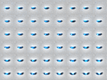
Bacteria duplicate their circular genome DNA by the progression of two replication forks bidirectionally. Each fork is driven by a multi-protein machinery named replisome that unzips, duplicates, and proofreads the DNA strands on-the-fly. Using time-lapse imaging, one can resolve 1 or 2 replisome foci across the whole cell cycle. However, given the asymmetry in foci intensity (e.g. bright and dim foci) or size/shape, it is challenging to detect the both replisomes with high accuracy and further provides the quantitative information of replisome positioning and mobility.
Details: The project will be focused on image processing and analysis using a synthetic or experimental data. The student will have to design a algorithm of tracking with a user-friendly GUI where a variety of functions are integrated, for example, the cell segmentation, spot detection and tracking, and etc.
This project is part of the EPFL Center for Imaging and in collaboration with the Laboratory of Experimental Biophysics (EPFL)
- Supervisors
- Daniel Sage, daniel.sage@epfl.ch, 021 693 51 89, BM 4.135
- Chen Zhang