Advanced machine learning for one-shot isotropic fetal MRI
2022
Master Diploma
Project: 00425
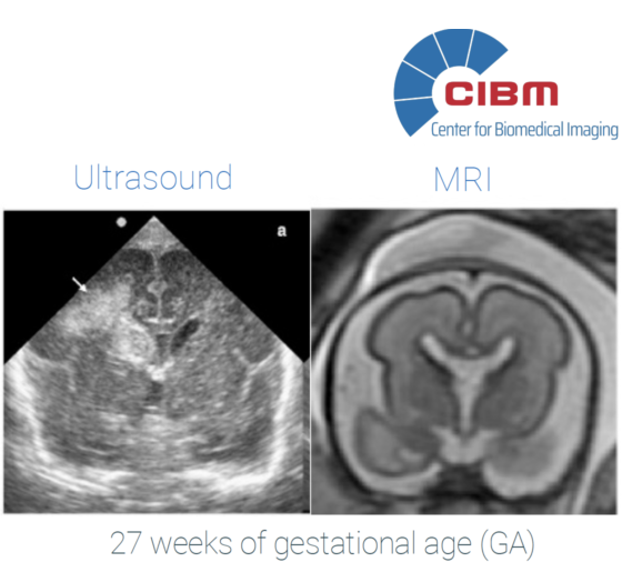
Description and objectives / summary: Magnetic resonance imaging (MRI) of the 3D structure of the foetal brain as it
develops inside the womb provides a high-resolution view of the brain's development. The resulting accuracy
is well beyond that achieved by ultrasound imaging, informs critical clinical diagnosis and prognosis, and
opens a path towards in-vivo quantitative studies. The capture of these MRI data, however, is challenging
to achieve. To mitigate imaging artifacts due to both fetal and mother motion, the MRI sequences have to be fast,
and resolution is typically compromised in one of the three axes. This is usually compensated by acquiring
separate stacks in which the low-resolution axis is alternated and blending the data using super-resolution techniques.
In this project, we aim to incorporate advances from deep neural networks and sparse representations to obtain the best isotropic resolution possible from a single MRI acquisition. These techniques leverage that the expected textures and patterns in an image are no different in the low-resolution dimension than they are in the high-resolution ones. Similar techiques have been previously applied to bio-imaging modalities where one of the three dimensions is also acquired at a lower resolution.
This project will be carried out at the CIBM Center for Biomedical Imaging, SP EPFL Mathematical Imaging Section, in collaboration with the SP CHUV-UNIL Computational Neuroanatomy & Fetal Imaging Section (Dr. Meritxell Bach Cuadra).
- Supervisors
- Pol del Aguila Pla, pol.delaguilapla@epfl.ch, BM 4.141
- Michael Unser, michael.unser@epfl.ch, 021 693 51 75, BM 4.136