Biomedical Imaging Group
Logo EPFL
    • Splines Tutorials
    • Splines Art Gallery
    • Wavelets Tutorials
    • Image denoising
    • ERC project: FUN-SP
    • Sparse Processes - Book Preview
    • ERC project: GlobalBioIm
    • The colored revolution of bioimaging
    • Deconvolution
    • SMLM
    • One-World Seminars: Representer theorems
    • A Unifying Representer Theorem
Follow us on Twitter.
Join our Github.
Masquer le formulaire de recherche
Menu
BIOMEDICAL IMAGING GROUP (BIG)
Laboratoire d'imagerie biomédicale (LIB)
  1. School of Engineering STI
  2. Institute IEM
  3.  LIB
  4.  Student Projects
  • Laboratory
    • Laboratory
    • Laboratory
    • People
    • Jobs and Trainees
    • News
    • Events
    • Seminars
    • Resources (intranet)
    • Twitter
  • Research
    • Research
    • Researchs
    • Research Topics
    • Talks, Tutorials, and Reviews
  • Publications
    • Publications
    • Publications
    • Database of Publications
    • Talks, Tutorials, and Reviews
    • EPFL Infoscience
  • Code
    • Code
    • Code
    • Demos
    • Download Algorithms
    • Github
  • Teaching
    • Teaching
    • Teaching
    • Courses
    • Student projects
  • Splines
    • Teaching
    • Teaching
    • Splines Tutorials
    • Splines Art Gallery
    • Wavelets Tutorials
    • Image denoising
  • Sparsity
    • Teaching
    • Teaching
    • ERC project: FUN-SP
    • Sparse Processes - Book Preview
  • Imaging
    • Teaching
    • Teaching
    • ERC project: GlobalBioIm
    • The colored revolution of bioimaging
    • Deconvolution
    • SMLM
  • Machine Learning
    • Teaching
    • Teaching
    • One-World Seminars: Representer theorems
    • A Unifying Representer Theorem

Students Projects

Proposals  On-Going  Completed  

Texture analysis for tumor histology images

Autumn 2005
Master Semester Project
Project: 00106

00106
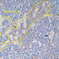
The assessment of drugs for cancer treatment is an important task to combat these diseases. A possible way is to analyze histology images of tumors, which look as shown in the figure. Image analysis is necessary to judge whether the drug treatment has significantly reduced the tumor proliferation. The two most important elements are the cell and vascular densities. This project consists of developping an ImageJ plugin that assists into the classification of tumor histology images. The first part is the measurement of the density of the cells, which appear as blue dots. Secondly, the method also needs to take into consideration the texture of the brown background which originates from blood vains.
  • Supervisors
  • Anonymous
  • Anonymous
  • Laboratory
  • Research
  • Publications
  • Code
  • Teaching
    • Courses
    • Student projects
Logo EPFL, Ecole polytechnique fédérale de Lausanne
Emergencies: +41 21 693 3000 Services and resources Contact Map Webmaster email

Follow EPFL on social media

Follow us on Facebook. Follow us on Twitter. Follow us on Instagram. Follow us on Youtube. Follow us on LinkedIn.
Accessibility Disclaimer Privacy policy

© 2025 EPFL, all rights reserved