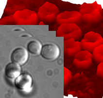
Digital quantitative phase recovery from focal series of brightfield microscopy images
2008
Master Semester Project
Project: 00162

Many biological specimens of interest to microscopists are inherently transparent and can only be visualized if they are stained. When staining the specimen is not an option, a specimen's phase structure can be visualized by techniques such as phase contrast (PC) microscopy or differential interference contrast (DIC) microscopy. The intensity distribution obtained via these techniques does generally not provide quantitative phase information, however.
In recent years, several new computational methods for quantitative phase recovery in brightfield microscopy have been proposed. They yield highly promising results and have the significant advantage of being compatible with any brightfield setup. The aim of this project is to review the state of the art of these methods and to propose an efficient implemention thereof in the form of a Java plug-in for ImageJ.
This algorithm could be applied to help the segmentation and the tracking of yeast cells.
Collaboration: Prof. Sebastien Maerkl, Life Science Faculty, EPFL.
- Supervisors
- François Aguet, francois.aguet@epfl.ch, 021 693 51 42, BM 4.141
- Michael Unser, michael.unser@epfl.ch, 021 693 51 75, BM 4.136