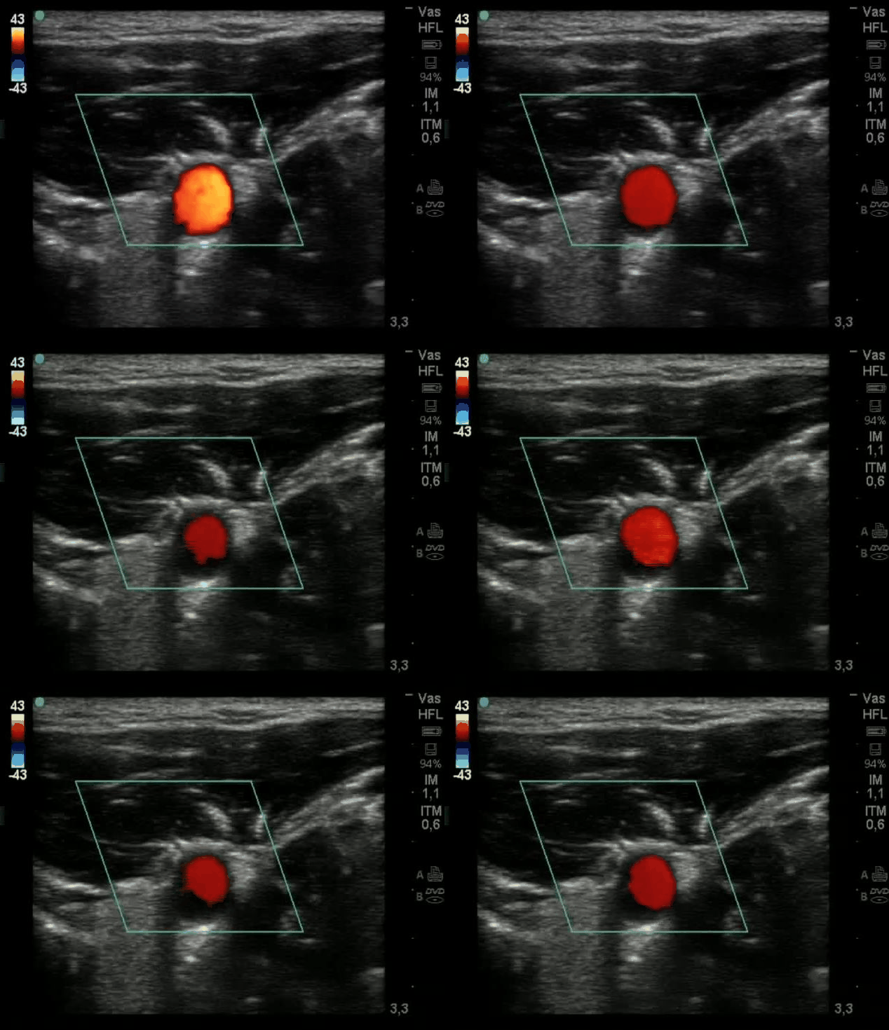
Segmentation of Doppler ultrasound images for monitoring the blood flow
Spring 2015
Master Semester Project
Project: 00289

The measurement and monitoring of vascular flow parameters are important in clinical medicine. The project consists in the development of an "active contour" (a.k.a. "snakes") model in order to obtain real-time measurements of the vascular flow (i.e. Doppler velocity x area) in Doppler ultrasound images. These images allow a colored visualization of the blood flow. A pre segmentation step consists in applying image processing and image analysis methods on the data. This work is part of a larger project whose goal is a clinical application and will be done in collaboration with the Department of Anaesthesiology at University Hospital Geneva (HUG) (collaboration with Raoul Schorer). The algorithm will be developped in JAVA and experiments will be done to define the application limits of the measurement method.
- Supervisors
- Anaïs Badoual, anais.badoual@epfl.ch, 31136, BM 4142
- Michael Unser, michael.unser@epfl.ch, 021 693 51 75, BM 4.136
- Daniel Sage, Raoul Schorer