Tracking Keratinocytes in co-culture
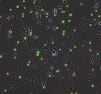
In practice, a biopsy of a healthy portion of the patients is made, and the keratinocytes it contains are grown on a feeder layer composed of cells called fibroblasts. A few weeks later, these stem cells give rise to continuous sheets of epidermis that can be transplanted back to the patient. Through cellular expansion, keratinocytes interact with the feeder layer and can undergo multiple fates: rest, symmetric division, asymmetric division, death and differentiation. Understanding the dynamics of keratinocytes regarding these features is important to improve skin transplantation techniques such as to incorporate hair follicles and natural mechanistic properties to the engineered skin. The goal of this project is to develop algorithms for identifying and tracking keratinocytes in co-culture- in the presence of other cells in label-free phase contrast and/or digital holographic microscopy videos. The student will have to implement novel tracking approaches of cells whose shape and number vary over time. Then, we will develop analytic tools to obtain quantitative data: cell count, division time, death rate and more.
- Supervisors
- Virginie Uhlmann, virginie.uhlmann@epfl.ch, 021 693 1136, BM 4.142
- Michael Unser, michael.unser@epfl.ch, 021 693 51 75, BM 4.136
- Daniel Sage, daniel.sage@epfl.ch, BM 4.135, Tel: 021 693 51 89