Biomedical Imaging Group
Logo EPFL
    • Splines Tutorials
    • Splines Art Gallery
    • Wavelets Tutorials
    • Image denoising
    • ERC project: FUN-SP
    • Sparse Processes - Book Preview
    • ERC project: GlobalBioIm
    • The colored revolution of bioimaging
    • Deconvolution
    • SMLM
    • One-World Seminars: Representer theorems
    • A Unifying Representer Theorem
Follow us on Twitter.
Join our Github.
Masquer le formulaire de recherche
Menu
BIOMEDICAL IMAGING GROUP (BIG)
Laboratoire d'imagerie biomédicale (LIB)
  1. School of Engineering STI
  2. Institute IEM
  3.  LIB
  4.  Student Projects
  • Laboratory
    • Laboratory
    • Laboratory
    • People
    • Jobs and Trainees
    • News
    • Events
    • Seminars
    • Resources (intranet)
    • Twitter
  • Research
    • Research
    • Researchs
    • Research Topics
    • Talks, Tutorials, and Reviews
  • Publications
    • Publications
    • Publications
    • Database of Publications
    • Talks, Tutorials, and Reviews
    • EPFL Infoscience
  • Code
    • Code
    • Code
    • Demos
    • Download Algorithms
    • Github
  • Teaching
    • Teaching
    • Teaching
    • Courses
    • Student projects
  • Splines
    • Teaching
    • Teaching
    • Splines Tutorials
    • Splines Art Gallery
    • Wavelets Tutorials
    • Image denoising
  • Sparsity
    • Teaching
    • Teaching
    • ERC project: FUN-SP
    • Sparse Processes - Book Preview
  • Imaging
    • Teaching
    • Teaching
    • ERC project: GlobalBioIm
    • The colored revolution of bioimaging
    • Deconvolution
    • SMLM
  • Machine Learning
    • Teaching
    • Teaching
    • One-World Seminars: Representer theorems
    • A Unifying Representer Theorem

Students Projects

Proposals  On-Going  Completed  

Characterization of myelin structure using image analysis techniques

Summer 2024
Master Semester Project
Project: 00451

00451

Context
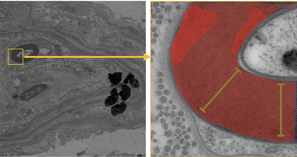
Room-temperature electron microscopy allows to study the ultrastructure of biological tissue and cells with nanometric resolution. In this project, this technique is used in particular to study the fine structure of myelinated fibers from healthy subjects and those diseased with a set of neurodegenerative diseases. In some myelinated fibers the structure is nicely preserved, while in some of them damage and swellings are visible in electron microscopy images. The goal of the project is quantify, analyze and categorize them according to their level of preservation using image analysis teachniques. This approach would be crucial to verify if myelin damage shows any correlation with the diseases.

Development
The goal of the project is to develop an image analysis workflow in Python to efficiently detect and segment all the myelinated fibers. This will enable to quantify the structure of the fibers and the damages areas in order to categorize the level of preservation. The methodology involves employing various image analysis techniques, such as denoising, edge and ridges detection, active contours, pixel classification, machine learning.

Requirement
Basic knowledge of signal or image processing Basic skills in coding

This project is a collaboration with the EPFL Center for Imaging and the Laboratory of Biological Electron Microscopy

  • Supervisors
  • Daniel Sage, daniel.sage@epfl.ch
  • Marta Di Fabrizio, marta.difabrizio@epfl.ch
  • Laboratory
  • Research
  • Publications
  • Code
  • Teaching
    • Courses
    • Student projects
Logo EPFL, Ecole polytechnique fédérale de Lausanne
Emergencies: +41 21 693 3000 Services and resources Contact Map Webmaster email

Follow EPFL on social media

Follow us on Facebook. Follow us on Twitter. Follow us on Instagram. Follow us on Youtube. Follow us on LinkedIn.
Accessibility Disclaimer Privacy policy

© 2025 EPFL, all rights reserved