Graphic STI
logo EPFL
text EPFL
english only
Biomedical Imaging Group
Research
BIG > Research > Applications Areas
CONTENTS

Home page

News & Events

Seminars

People

Research

Publications

Tutorials & Reviews

Recent Talks

Demos

Download Algorithms

Jobs and Trainees

Teaching

Student Projects

Intranet

Applications Areas


Medical Imaging
Medical Imaging

Medical imaging constitutes the primary area of application of our mathematical and signal-processing techniques. The important aspects that need to be considered are

  • the sophistication and variety of imaging techniques (MRI, CT, PET, ultrasound)—the design of optimized algorithms requires a thorough understanding of the image-formation process and of the various sources of distortion (noise, point-spread function);
  • the inter-disciplinary nature of the field: A close interaction with clinicians (radiologists) is essential for understanding what the important issues are. Medical experts are also required for establishing diagnostic criteria and for assessing the results;
  • the large amount of data that needs to be handled: Typical medical-imaging data are 3D—they can also be multimodal, and/or dynamic (3D + time);
  • the quest for quality and reproducibility: Medical images are extremely valuable—the implication of an incorrect diagnosis or analysis can be devastating;
  • the necessity for a careful validation of algorithms on clinical cases. This last point is crucial if one wants to have an impact on the field.

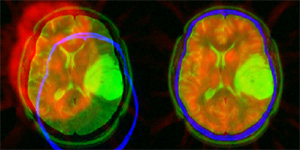
The current focus of our research is the interpolation of medical images, image reconstruction, 3D visualization, various types of image registration (e.g., intra- or inter-modal, elastic, 2D/3D), wavelet-based techniques, and motion estimation from echocardiograms. Our greatest impact so far has been the introduction of the spline methodology in medical imaging: Some of our high-quality interpolation algorithms have been incorporated in standard imaging software such as SPM.

Current research projects

Past research projects


Advanced Image Processing in Biology

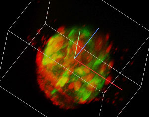
With the recent development of fluorescent probes and of new high-resolution microscopes (e.g., confocal, two-photon, FRET), biological imaging has grown quite sophisticated and is presently having a profound impact on the way research is being conducted in molecular biology. Biomedical scientists can visualize sub-cellular components and processes, both structurally and functionally, in two or three dimensions, at different wavelengths (spectroscopy), and can perform time-lapse imaging to investigate cellular dynamics. The data analysis and processing techniques that are currently used in the field, however, are still relatively crude if one compares them with the state-of-the art in medical imaging.

This motivates our present research effort which is to develop novel image-processing techniques to improve the quality of these images, to enhance specific features (e.g., spots, filaments), and to extract quantitative measurements from the data.

Some aspects that are specific to this type of imaging research are:

  • the sophistication and variety of imaging techniques;
  • the increasing need for quantitative image analysis;
  • challenging data that are often very noisy, and at the limit of resolution;
  • multiplicity of dimensions: 2D or 3D, time (dynamic imaging), multi-spectral;
  • modeling of the acquisition process: optical system (2D or 3D point-spread function), noise (photon-limited).

Current research projects

Past research projects

© 2022 EPFL • webmaster.big@epfl.ch • 11.08.2022